Background and clinical problems
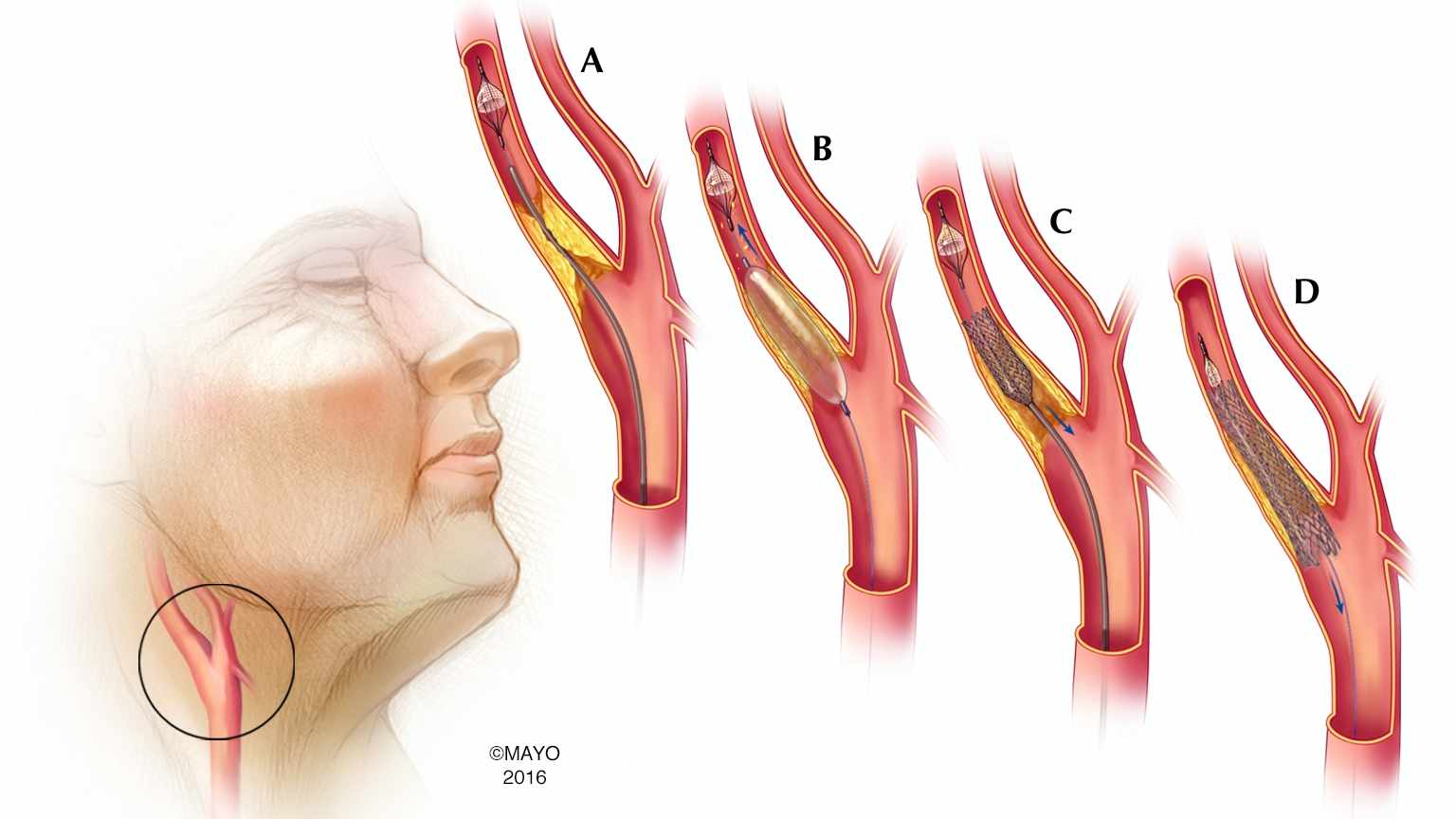
The DIVECAP-project focusses on two types of pathology of the extracranial carotid artery. The carotid arteries are the main arteries for blood supply to the brain. These arteries can be narrowed (stenosed) or widened (aneurysmatic). Both pathologies can cause brain damage by transient ischemic attack (TIA) or stroke. When symptoms are present, treatment is indicated.
One of the treatment options is endovascular intervention, also called carotid artery stenting (CAS). Technical improvements in CAS may reduce the stroke risk during and immediately after intervention. Then, CAS may become an equally effective treatment (compared to surgery) for carotid artery disease.
The degree of stenosis, important for the decision to treat or not to treat, is measured on Duplex ultrasound and computed tomography angiography (CTA) or magnetic resonance angiography (MRA). Recent research to inter- and intra-observer variability in determination of stenosis degree resulted in low accuracy and precision. Therefore, there is a need for a method to reproducibly measure stenosis degree.
Approaches
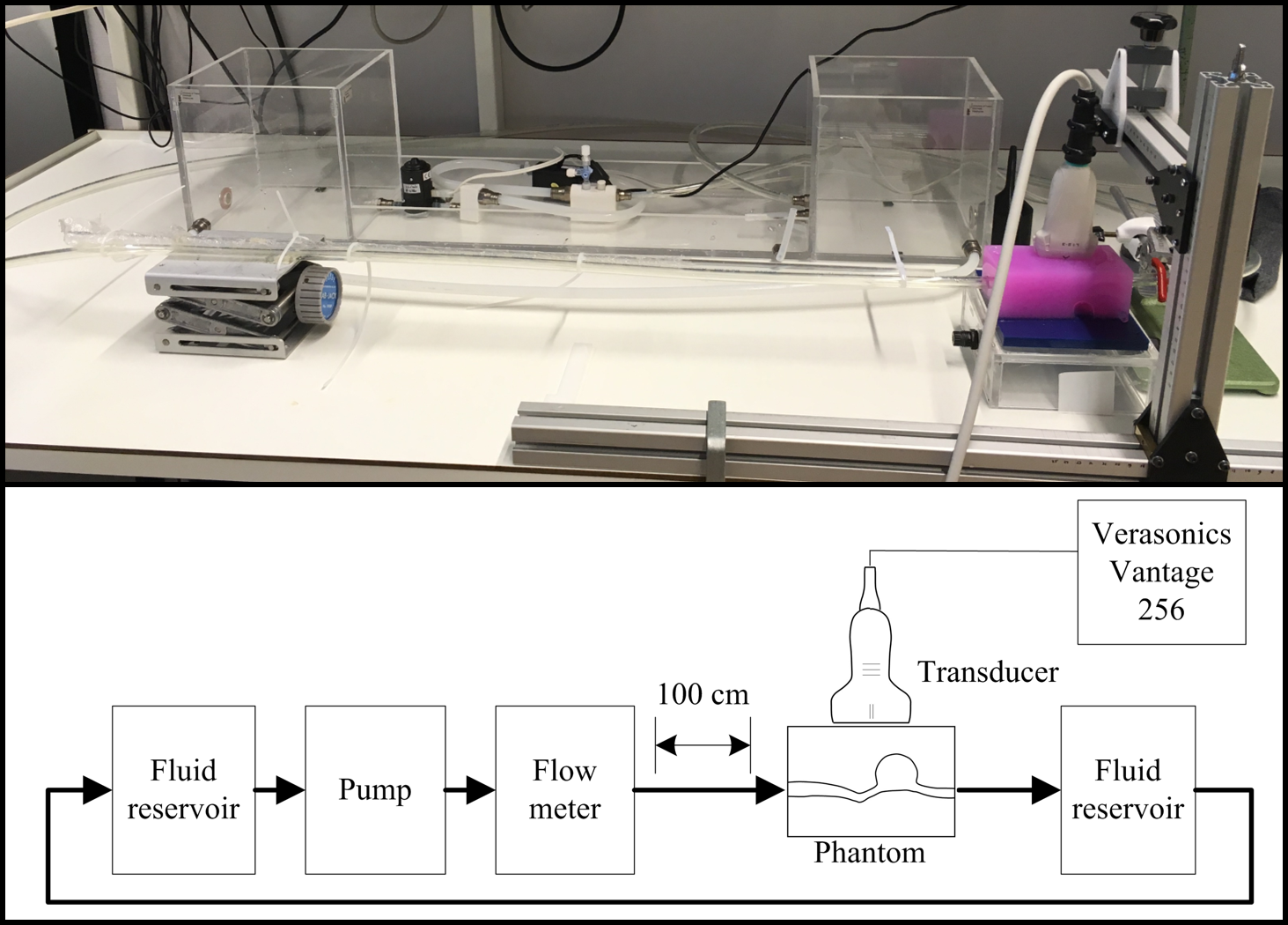
To study the effects of stent placement on the carotid blood flow, in vitro flow studies will be performed. The results can eventually be used for improvements in stent design and device development. A novel ultrasound technique, high-frame-rate contrast-enhanced ultrasound combined with particle image velocimetry (HFR echoPIV), will be implemented to study flow patterns in (patient-specific) carotid artery phantoms/models.
To improve stenosis degree measurements, objective analysis of the diseased carotid artery is desired. Therefore, this project focuses on automatic segmentation of the carotid artery. To obtain these segmentations, machine/deep learning techniques will be applied. Once the automatic segmentations are acquired, they will be used to explore novel and more accurate measurements for stenosis degree determination.
Topics
- Ultrasound imaging
- Blood flow studies
- Phantom/model fabrication
- 3D printing
- Computed tomography angiography
- Medical image segmentation
- Neural networks; machine/deep learning
Collaborations
- Vascular surgery department of University Medical Centre Utrecht (UMC Utrecht)
- Radiology department of hospital Ziekenhuis Groep Twente (ZGT) Hengelo/Almelo
If you are interested in a master or bachelor assignment, please contact Astrid Hoving (a.m.hoving@utwente.nl).