The MyoFLOW project aims to contribute to the validation, calibration and evaluation of quantitative myocardial perfusion imaging via phantom modeling.
Motivation
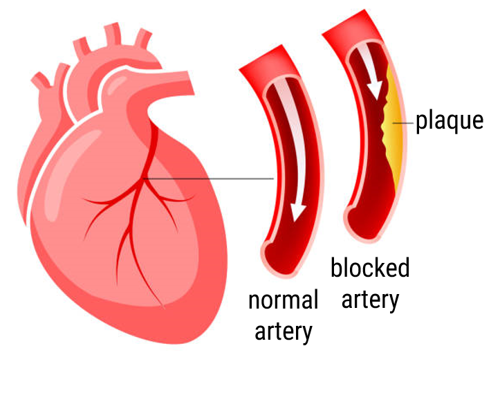
Plaque buildup inside the coronary arteries (arteries that supply the heart muscle) can cause a flow limitation towards the myocardial tissue (heart muscle tissue). If left untreated, this tissue could eventually die, leading to infarction. Proper medical decision-making is key, since coronary artery disease remains one of the most common causes of death in the western world.

Myocardial perfusion imaging is an important imaging technique to diagnose perfusion / blood flow defects. During such diagnostic test, a radio-active tracer is injected into a patient’s blood stream. Then the distribution of tracer inside the heart muscle is imaged over time using positron emission tomography (PET) or single photon computed tomography (SPECT) imaging. A reduced uptake in a certain region (relative to other regions) indicates a perfusion defect. However, evaluation of the resulting images is rather subjective and could underestimate a patient’s risk.
Blood flow quantification software, based on indicator dilution theory, could improve diagnostic accuracy and precision. Such software is tested extensively for PET imaging. However, can we also use it for clinical practice with SPECT scanners?
Topics
- Multimodal imaging
- Blood flow modeling / quantification
- Phantom development
- 3D printing
- Process control of cardiovascular flow circuit
- Clinical testing and validation
Collaborations
- Multi Modality Medical Imaging group (UT)
- Ziekenhuis Groep Twente hospital
- European Institute for Molecular Imaging Münster
If you are interested in a master or bachelor assignment related to the above mentioned topics, please contact Marije Kamphuis: m.e.kamphuis@utwente.nl.
Associated assignments
| Development of a myocardial perfusion phantom | Gijs de Vries |
